関西医科大学 医工学センター

Research Theme

Raman Spectroscopy: A Step Forward in Molecular Medicine

We investigate biological tissues, cells, microorganisms, and extracellular vesicles such as exosomes to elucidate molecular signals that emerge at the earliest stages of disease, with the aim of realizing early diagnosis and preventive intervention.

At the core of our research is Raman spectroscopy, a powerful analytical technique that enables non-destructive analysis of biological samples without the need for labeling or complex sample preparation. By visualizing molecular compositions and biochemical changes with high sensitivity, Raman spectroscopy offers great potential as a next-generation precision diagnostic technology.

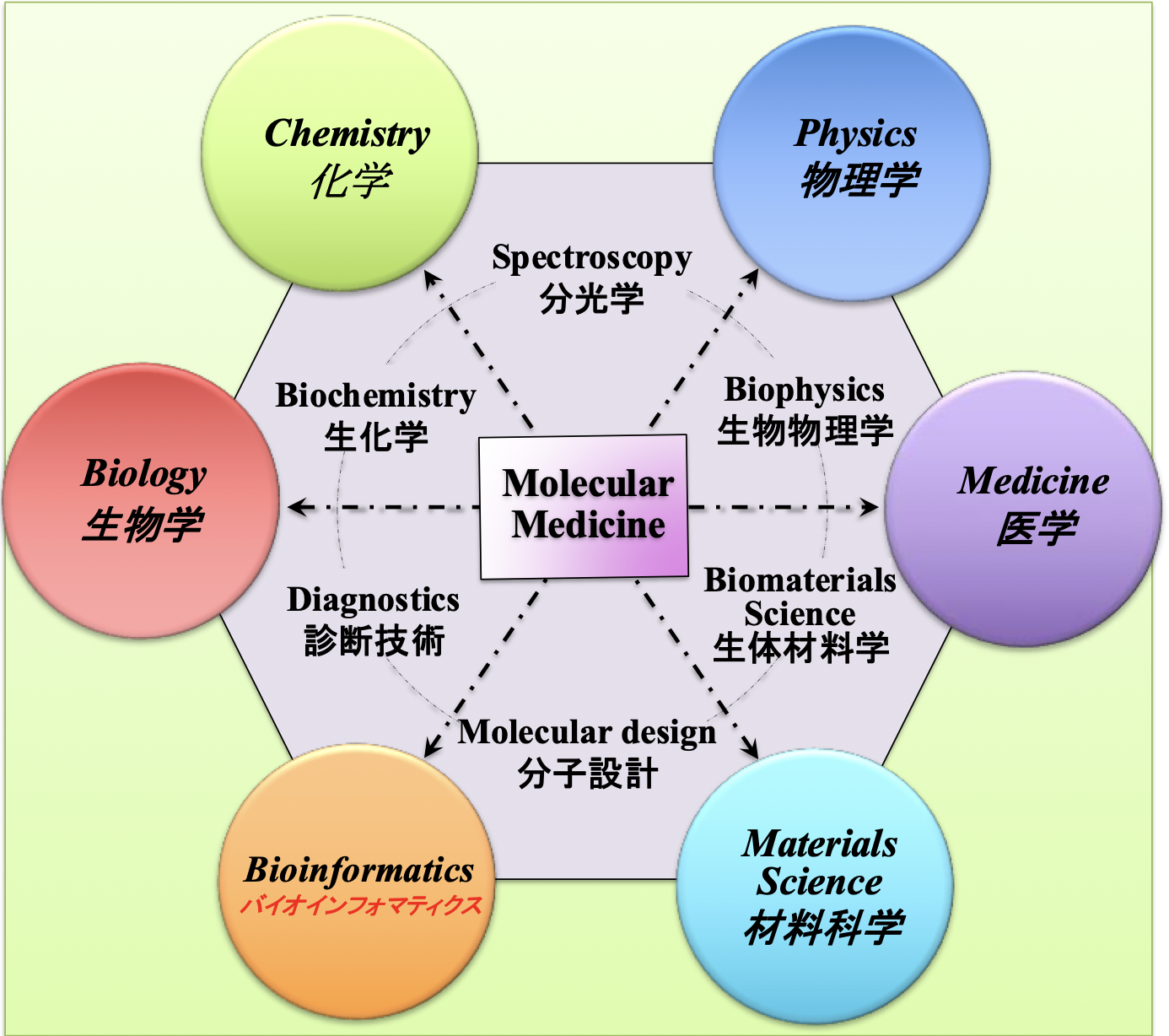
At our center, we integrate medicine and engineering—including physics, chemistry, and data science—to transform molecular information into clinically meaningful insights. Through this interdisciplinary approach, we aim to open new frontiers in preventive medicine and precision healthcare.


Research Contents

Raman signatures of type A and B influenza viruses: molecular origin of the “catch and kill” inactivation mechanism mediated by micrometric silicon nitride powder

Journal: RSC Chemical Biology
Published: 1 February 2025
https://doi.org/10.1039/D4CB00237G

Raman Fingerprints of SARS-CoV-2 Omicron Subvariants: Molecular Roots of Virological Characteristics and Evolutionary Directions

Journal: ACS infectious Diseases
Published: October 2023
https://doi.org/10.1021/acsinfecdis.3c00312

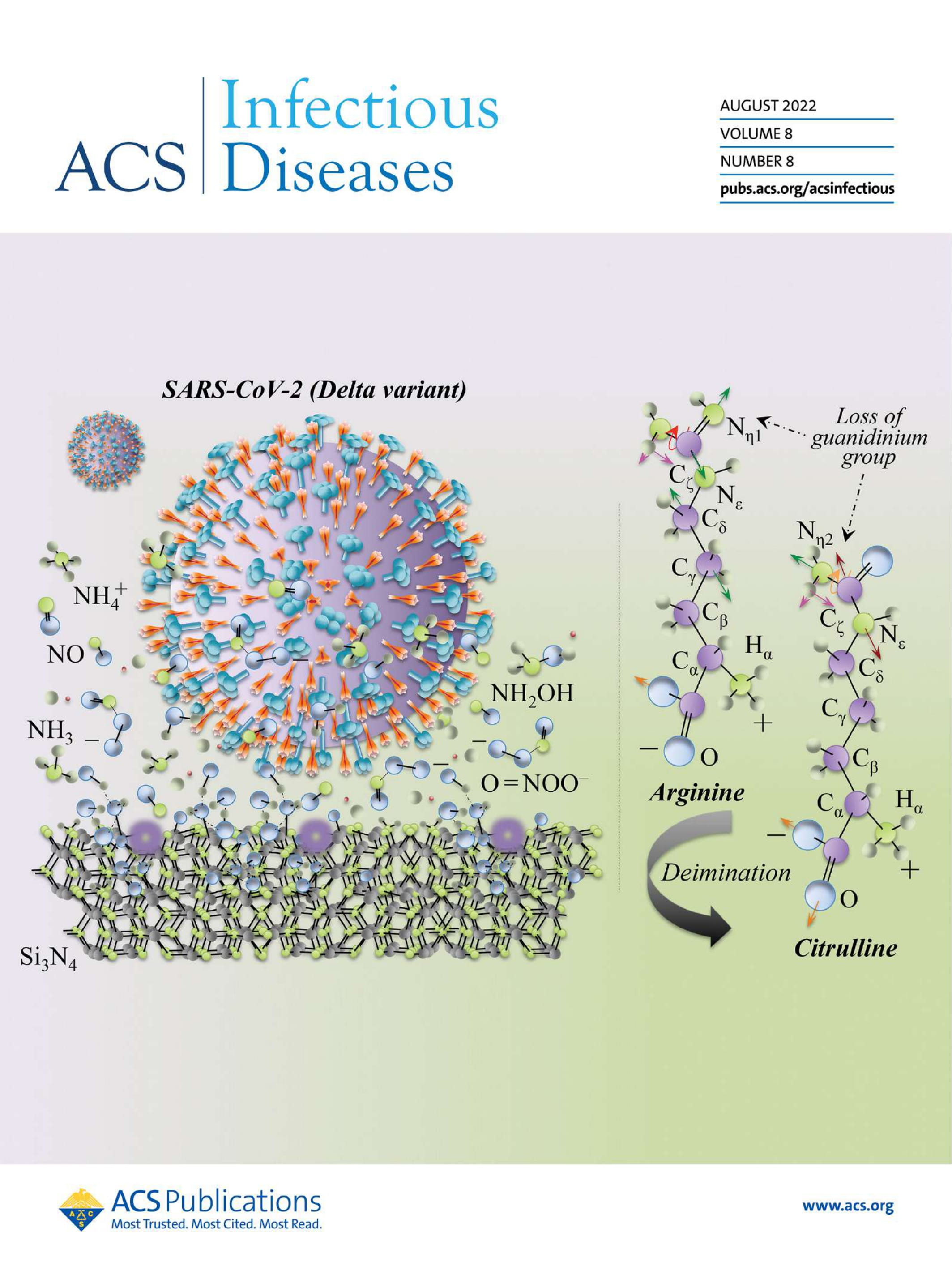
Raman Fingerprints of the SARS-CoV-2 Delta Variant and Mechanisms of Its Instantaneous Inactivation by Silicon Nitride Bioceramics

Journal: ACS Infectious Diseases
Published: August 2022
https://doi.org/10.1021/acsinfecdis.2c00200

Incorporating Si3N4 into PEEK to Produce Antibacterial, Osteoconductive, and Radiolucent Spinal Implants

Journal: Macromol. Biosci
Published: June 2018
https://doi.org/10.1002/mabi.201800033

Raman spectroscopy of piezoelectrics

Journal: Journal of Applied Physics
Published: June 2013
https://doi.org/10.1063/1.4803740

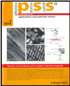
Spatially resolved Raman and cathodoluminescence probes in electronic materials: Basics and application

Journal: physica status solidi (a)
Published: May 2011
https://doi.org/10.1002/pssa.201000785

On the role of oxygen vacancies and lattice strain in the tetragonal to monoclinic transformation in alumina/ zirconia composites and improved environmental stability

Journal: Biomaterials
Published: September 2010
https://doi.org/10.1016/j.biomaterials.2010.05.035

Raman tensor analysis of sapphire single crystal and its application to define crystallographic orientation in polycrystalline alumina

Journal: physica status solidi (b)
Published: August 2009
https://doi.org/10.1002/pssb.200945137

Raman piezo-spectroscopic analysis of natural and synthetic biomaterials

Journal: Analytical and Bioanalytical Chemistry
Published: October 2004
https://link.springer.com/article/10.1007/s00216-004-2780-1

1. Raman spectroscopy in cell biology and microbiology

Journal: Journal of Raman Spectroscopy
Published: April 2021
https://analyticalsciencejournals.onlinelibrary.wiley.com/doi/epdf/10.1002/jrs.6204

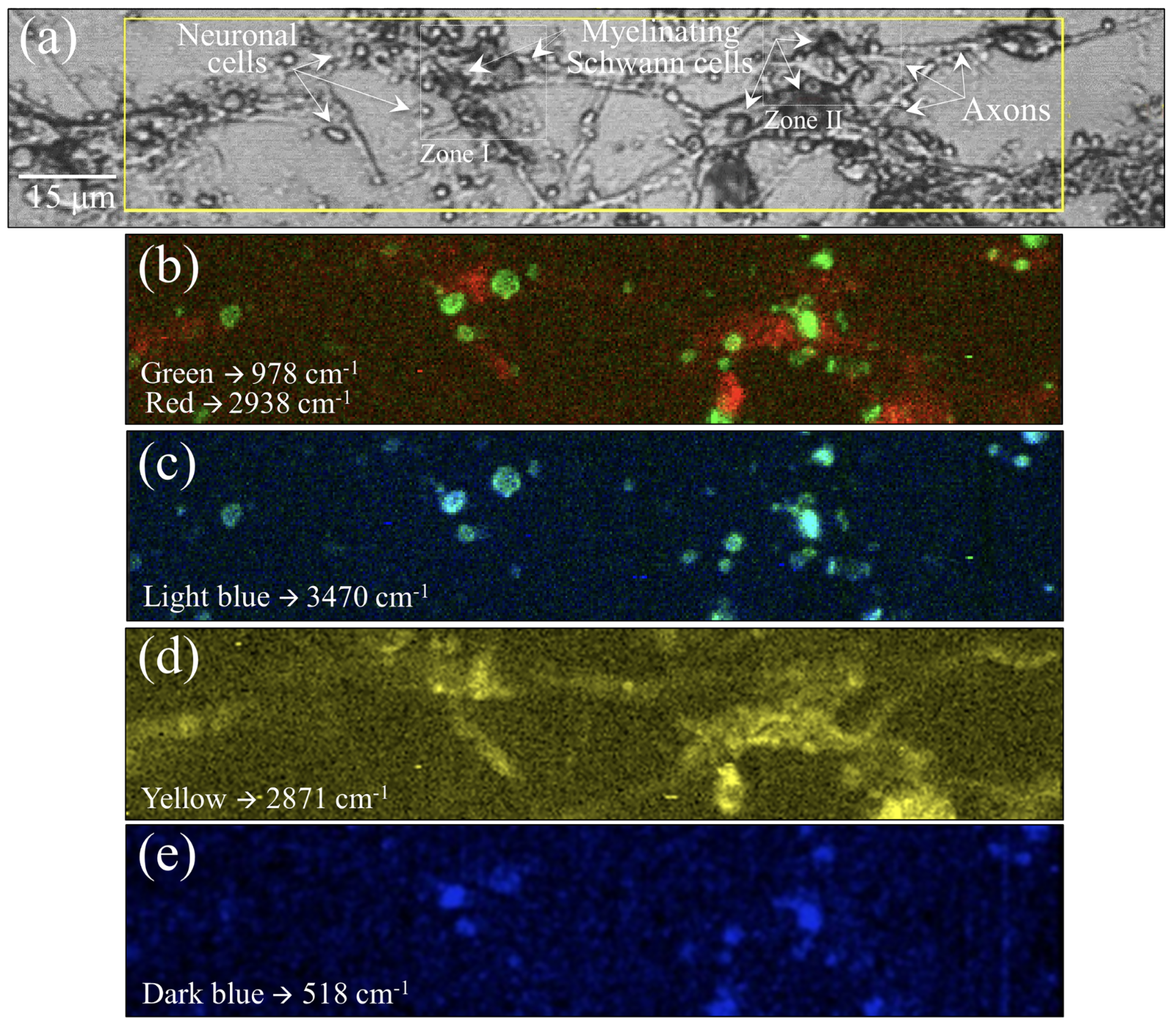
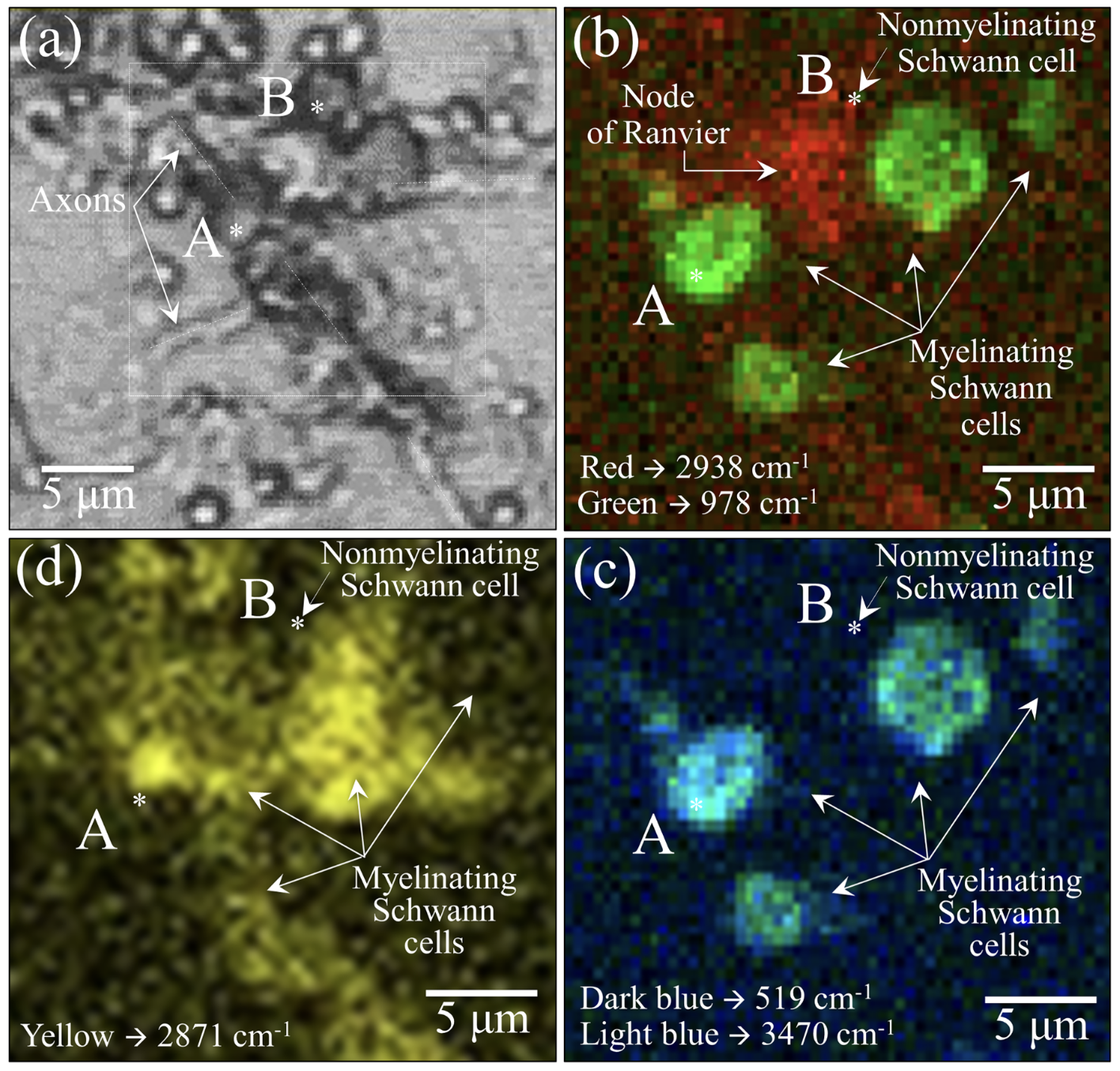
Raman imaging of living PC12/IFRS1 cell coculture (a). Maps taken at the Raman wavenumbers of 978 and 2938 cm-1 are merged in green and red, respectively (b), those taken at 519 and 3470 cm-1 are in dark and light blue, respectively (c), and that at 2971 cm-1 is in yellow (d). Imaging enables to visualize the extent of myelination in individual cells and the chemical events behind such behavior.

2. Oxysulfur and organosulfur toxins: the hidden mediators of the oral-systemic connection

Journal: Frontiers in Microbiology
Published: June 2026
https://doi.org/10.3389/fmicb.2026.1823575

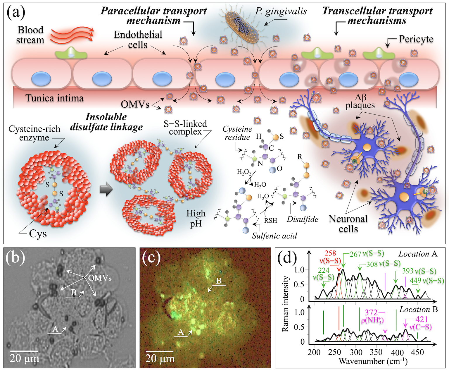
(a) Schematic draft of the possible routes by which P. gingivalis OMVs could penetrate the BBB, contaminate the brain tissue, and degrade neuronal cells; also in (a), draft showing the formation of insoluble disulfate linkages at enzymatic sites and at oxidized cysteine sites, ultimately leading to plaque formation. Optical image (b) and hyperspectral Raman map (c) collected on a live neuronal cells culture contaminated with micrograms of P. gingivalis OMVs; the Raman map represents Raman intensities at two specific wavenumbers: 258 cm-1 (red) and 267 cm-1 (green), representing S–S bond stretching in dithionite and tetrathionate molecules, respectively. The overlap of the two colors is seen in yellow; (d) shows local Raman spectra extracted from the map at the two selected locations A and B (cf. labels in inset). The abbreviation Aβ stands for amyloid-β.

3. Raman signatures of Cnm-positive Streptococcus mutans: I, the molecular origin of cerebral microbleeds

Journal: Frontiers in Microbiology
Published: April 2026
https://doi.org/10.3389/fmicb.2026.1784125

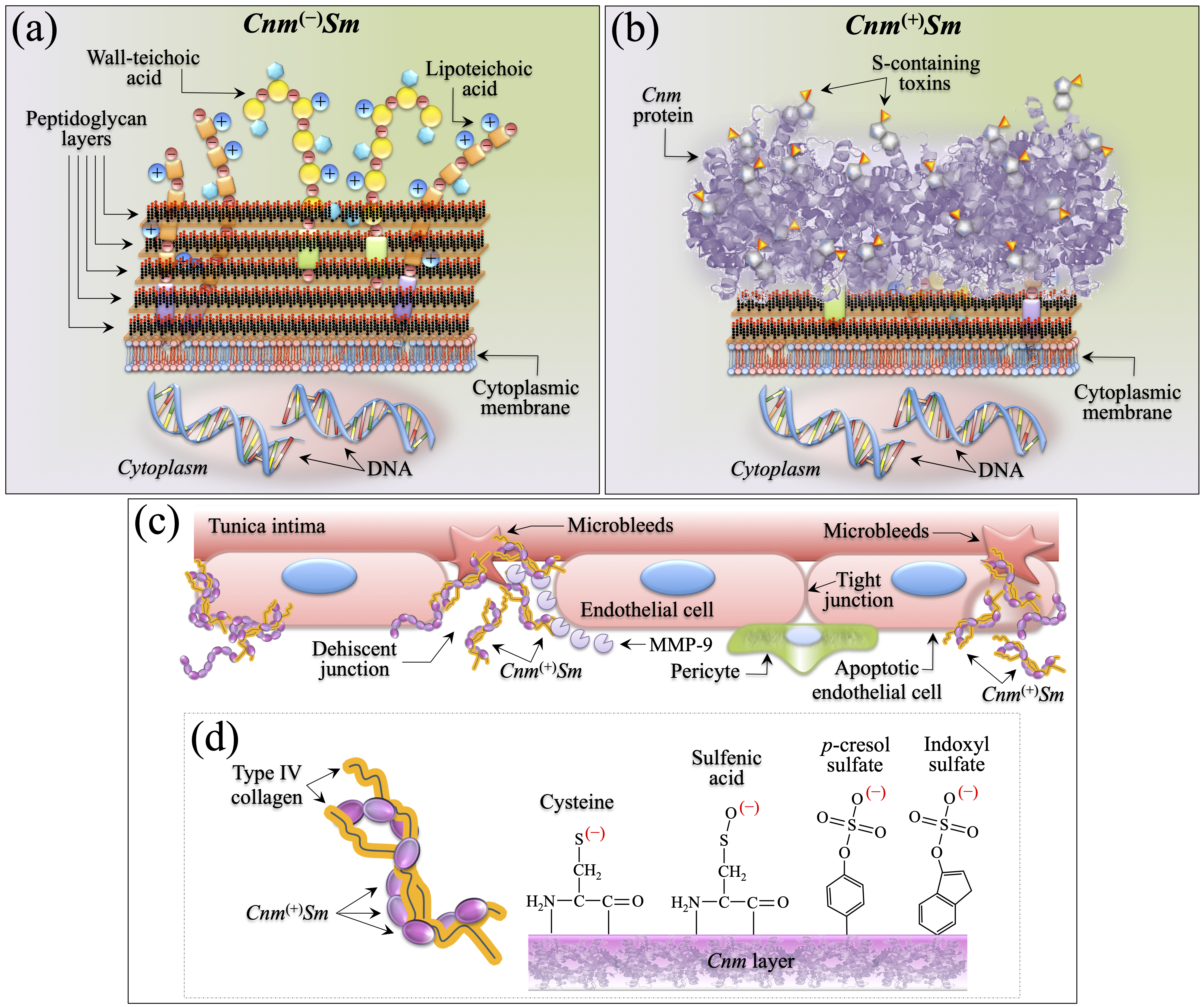
Drafts summarizing the main structural differences between Cnm(−)Sm(a) and Cnm(+)Sm(b) bacterial cells; (a) shows a structure rich in peptidoglycans, wall-teichoic and lipoteichoic acids (both composed of a linkage, repeat, phosphate, D-alanylation, and glycosylation units), while (b) launch a highly disordered Cnm protein layer wrapping the cell and a significant simplification of the peptidoglycan wall-structure (including teichoic and lipoteichoic moieties). The highly disordered Cnm protein in (b) is replenished of toxins exhibiting sulfenic and sulfonic acid termini. In (c), a draft is offered of the mechanism by which blood-circulating Cnm(+)Sm bacteria causes intracerebral hemorrhage: binding to denuded basement membranes at dehiscent cellular junctions (left side) and direct invasion of endothelial cells (right side). Collagen-adhesion characteristics through sulfur chemistry are depicted in (d): direct attachment to type IV collagen through S–S bonding by cysteine and sulfenic acid to indirect pathways including hyperglycemia-inflammation-coagulation induced by indoxyl sulfate and p-cresol sulfate moieties.

4. Raman Metabolomics of Candida auris Clades: Profiling and Barcode Identification

Journal: International Journal of Molecular Sciences
Published: October 2022
https://doi.org/10.3390/ijms231911736

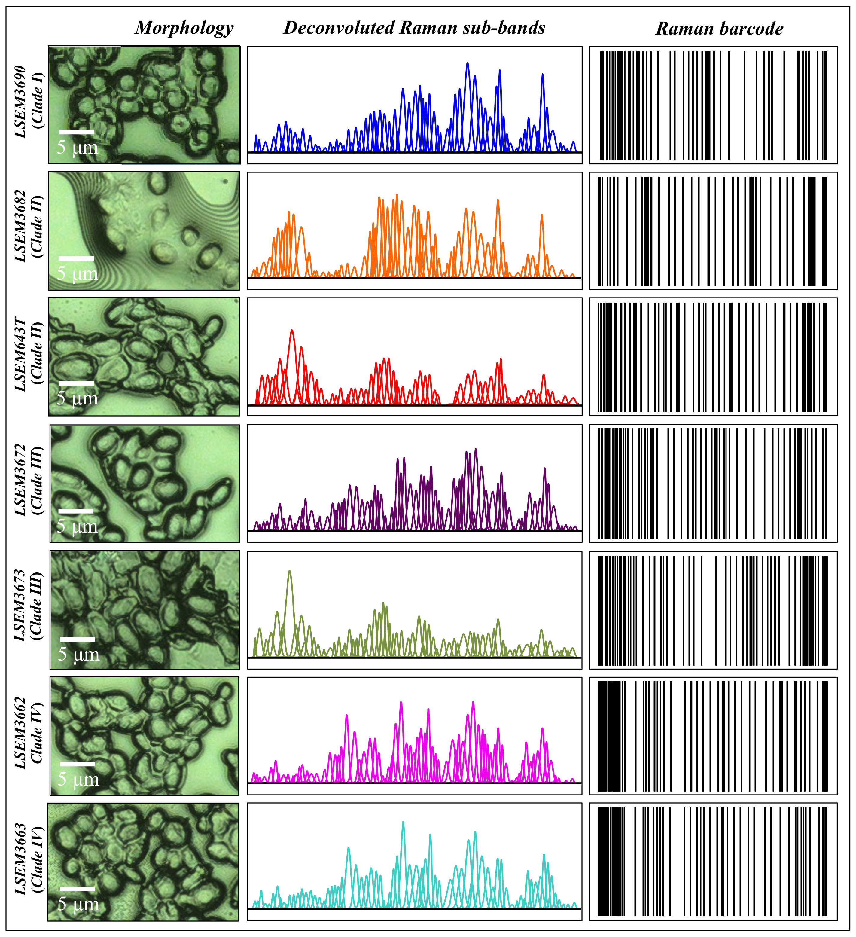
From left to right: high-resolution micrographs of different C. auris clades/subclades (cf. labels in inset), series of Gaussian–Lorentzian subclades (i.e., the same as those shown in the deconvoluted spectra of Figure 2) obtained from a machine-learning-based spectral deconvolution (cf. Section 2.3) for each sample, and Raman barcodes.

5. Raman Fingerprints of Rice Nutritional Quality: A Comparison between Japanese Koshihikari and Internationally Renowned Cultivars

Journal: Foods
Published: November 2021
https://doi.org/10.3390/foods10122936

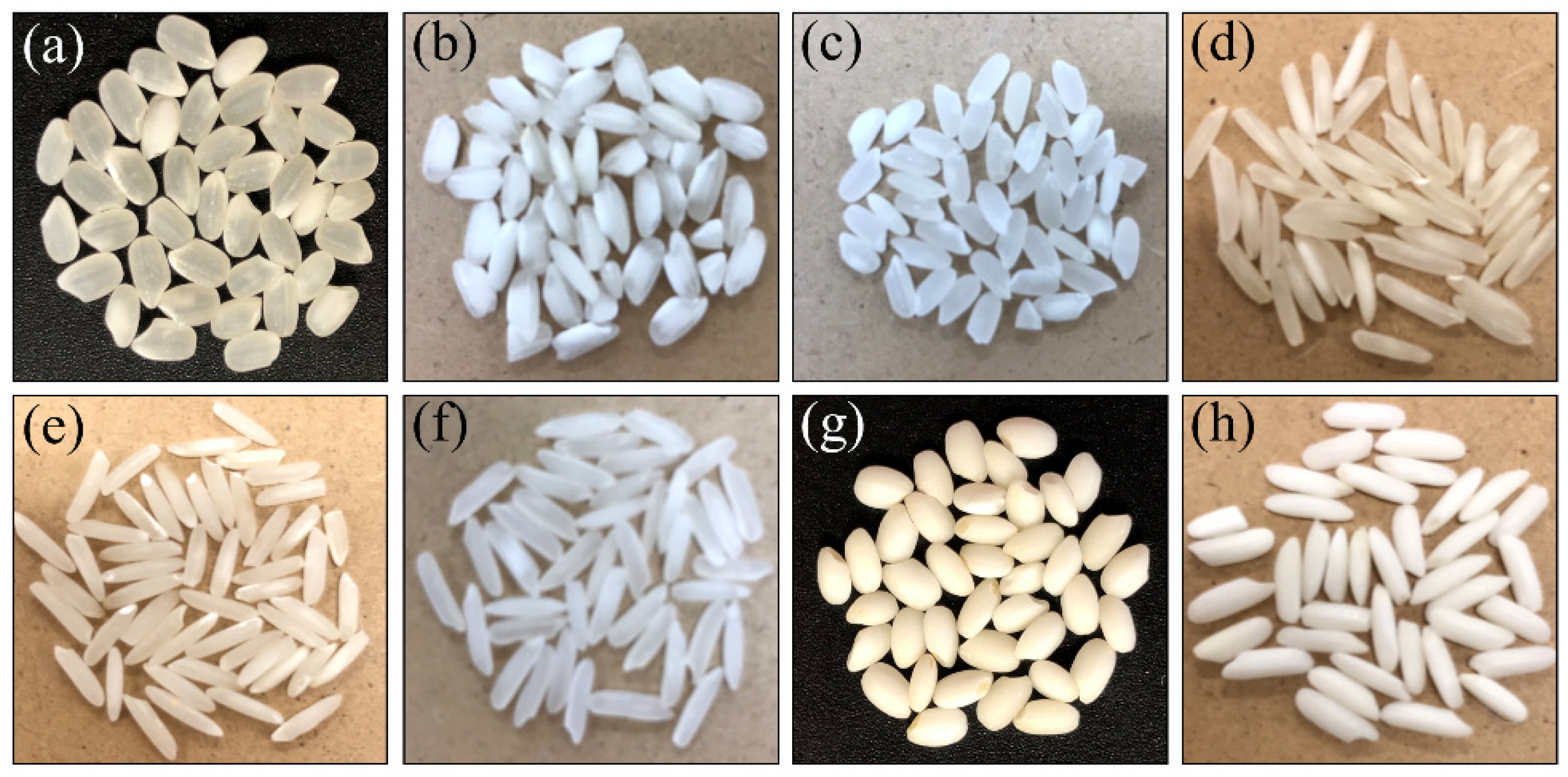
Left side: Photographs of (a) Koshihikari (Japan), (b) Carnaroli (Italy), (c) Calrose (USA), (d) Basmati (India), (e) Basmati (Pakistan), (f) Jasmine (Thailand), (g) Mochigome (Japan) and (h) Mochigome (Thailand).
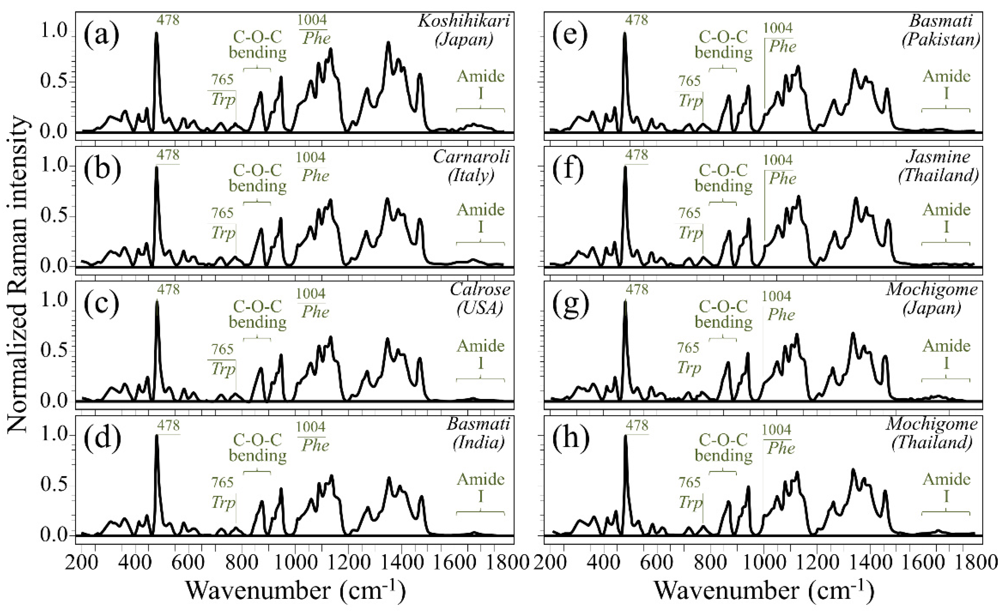
Right side: Raman spectra collected on as-received kernels of the above eight different rice cultivars (cf. labels in inset) in the spectral region 200–1800 cm-1; the spectra are averaged over 30 spectra per each type of cultivar and normalized to the glucose ring stretching band at ~478 cm-1 (cf. label in inset). The abbreviations Phe and Trp refer to phenylalanine and tryptophan, respectively, while the wavenumbers in inset are given in cm-1 units.

6. Silicon Nitride: A Bioceramic with a Gift

Journal: ACS Applied Materials & Interfaces
Published: June 2019
https://doi.org/10.1021/acsami.9b07997

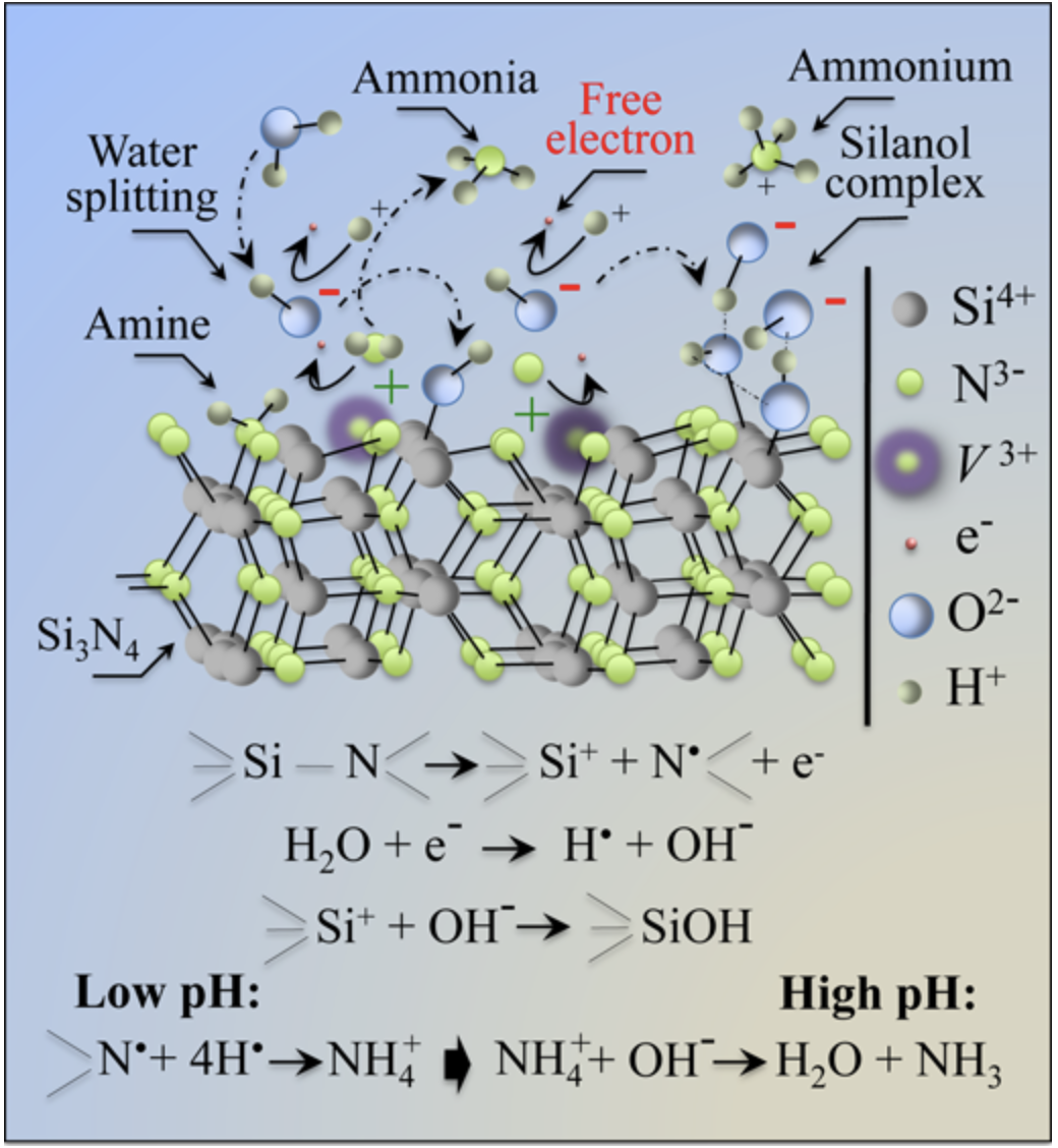
Draft of the surface chemistry of silicon nitride (Si3N4) bioceramics, which concurrently confers to this biomaterial both osteogenic and antibacterial properties.

Research Facilities

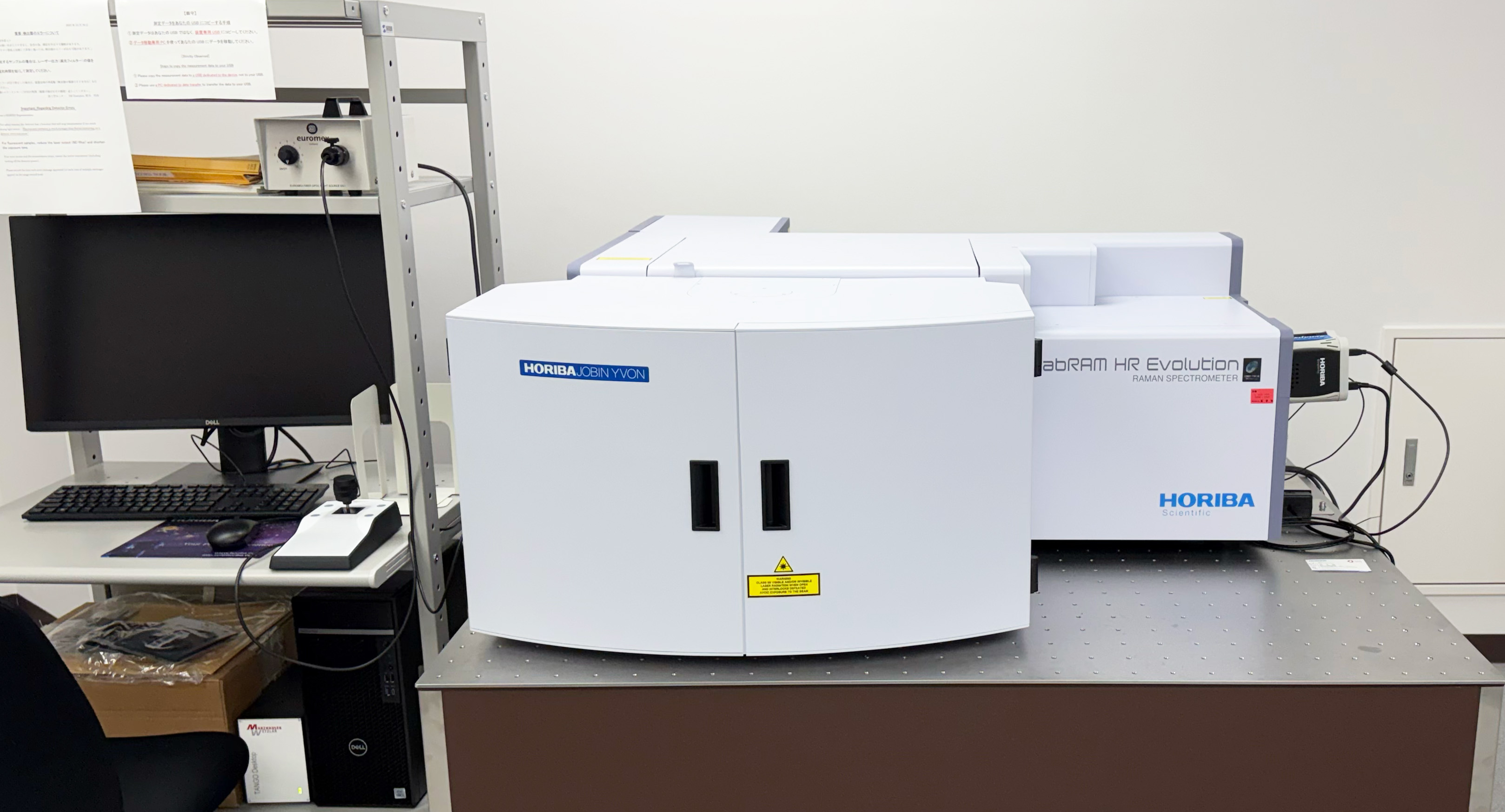
LabRAM HR Evolution RAMAN SPECTROMETER
(HORIBA Ltd.)

This Raman spectrometer offers advanced confocal imaging capabilities in both 2D and 3D, ideal for micro and macro measurements. It provides high-resolution confocal Raman microscopy, thus ensuring detailed analysis with speed and confidence.

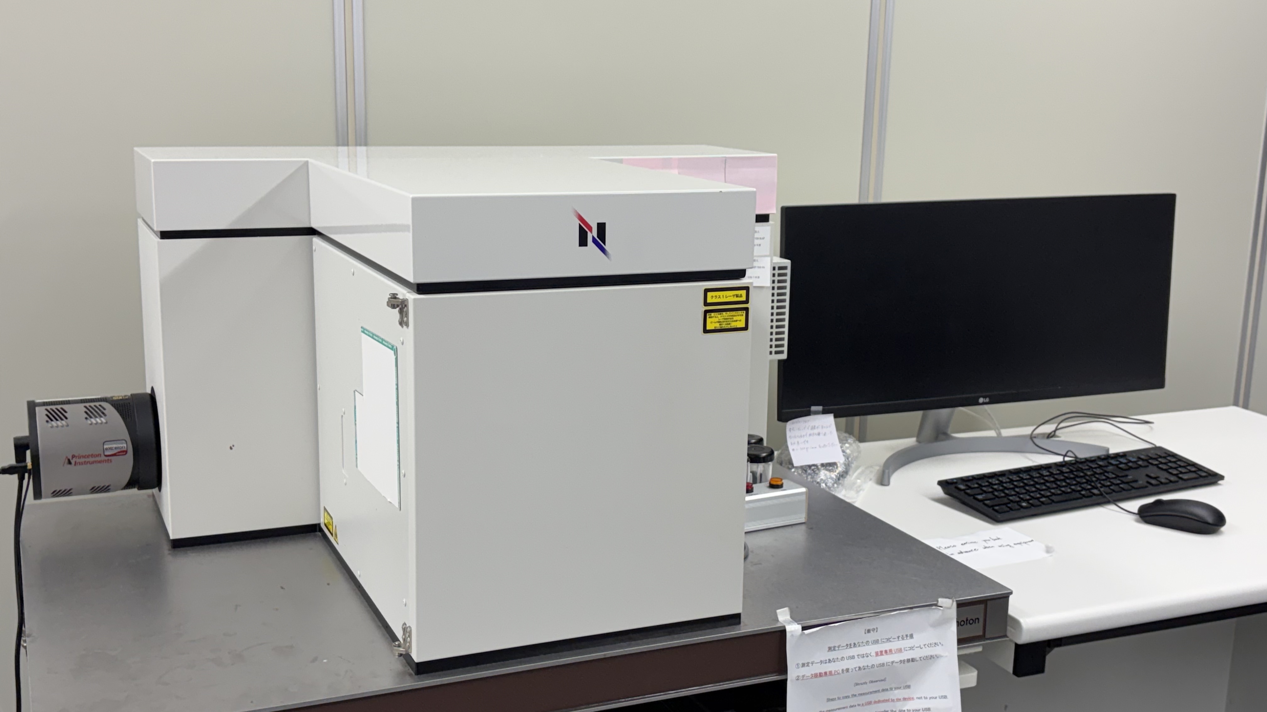
RAMANtouch (Nanophoton Corp.)

The World's Fastest and Highest-Resolution Laser Raman Image Microscope Equipped with a newly designed spectrometer and optical components that incorporate cutting-edge optical technology.

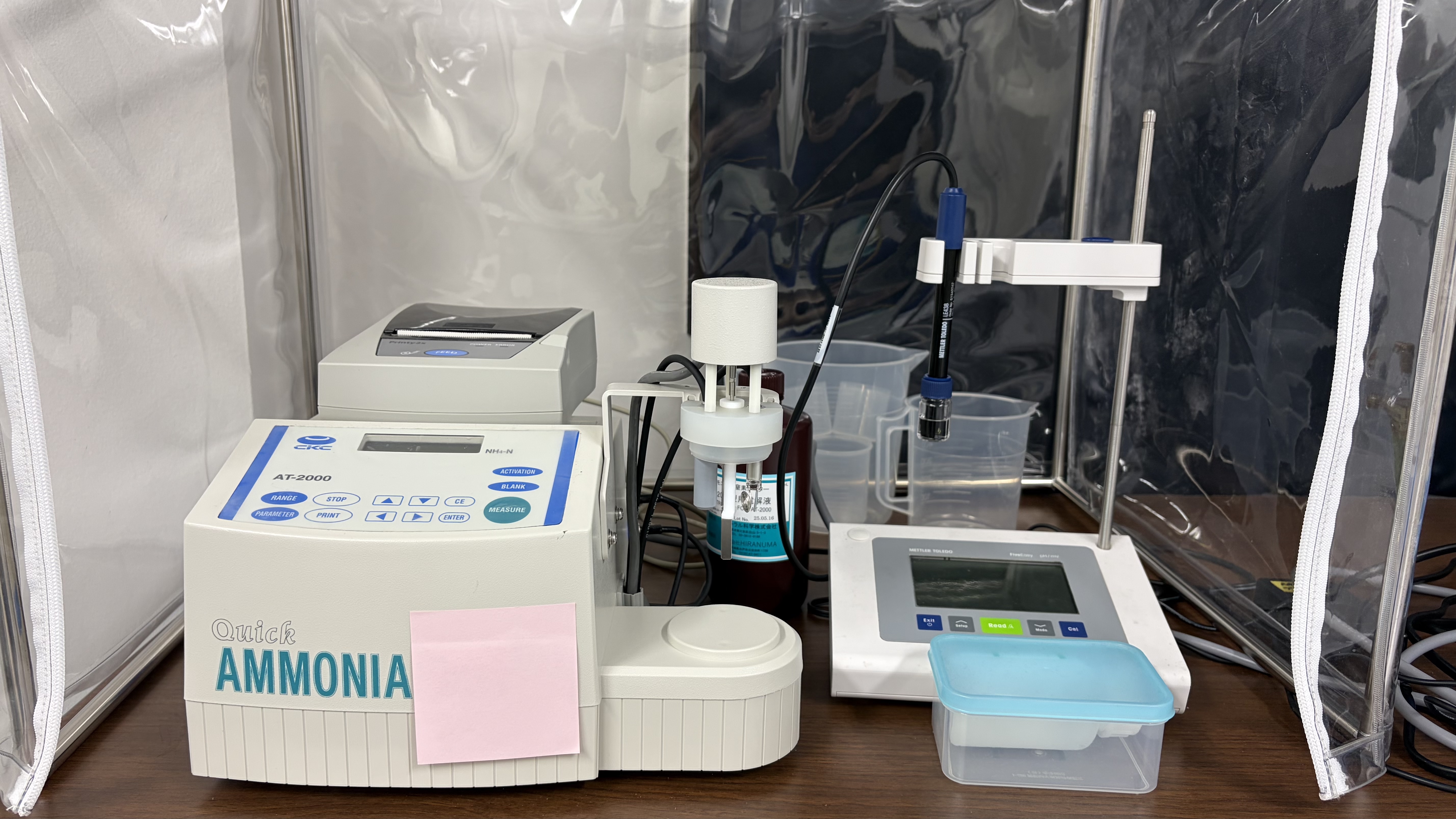
Ammonia Measurement by Coulometric Method - Quick Ammonia AT-2000 Model

Rapid, accurate, and easy measurement of ammonia suitable for a wide range of applications, from freshwater to seawater.